
AutoBrachy · AI-Powered GYN HDR Brachytherapy Planning
A draft plan for the physicist, in minutes
AutoBrachy digitizes the applicator, contours OARs and the target, and optimizes dwell times from the planning CT — producing a draft plan your physicist reviews, edits, and approves before treatment.
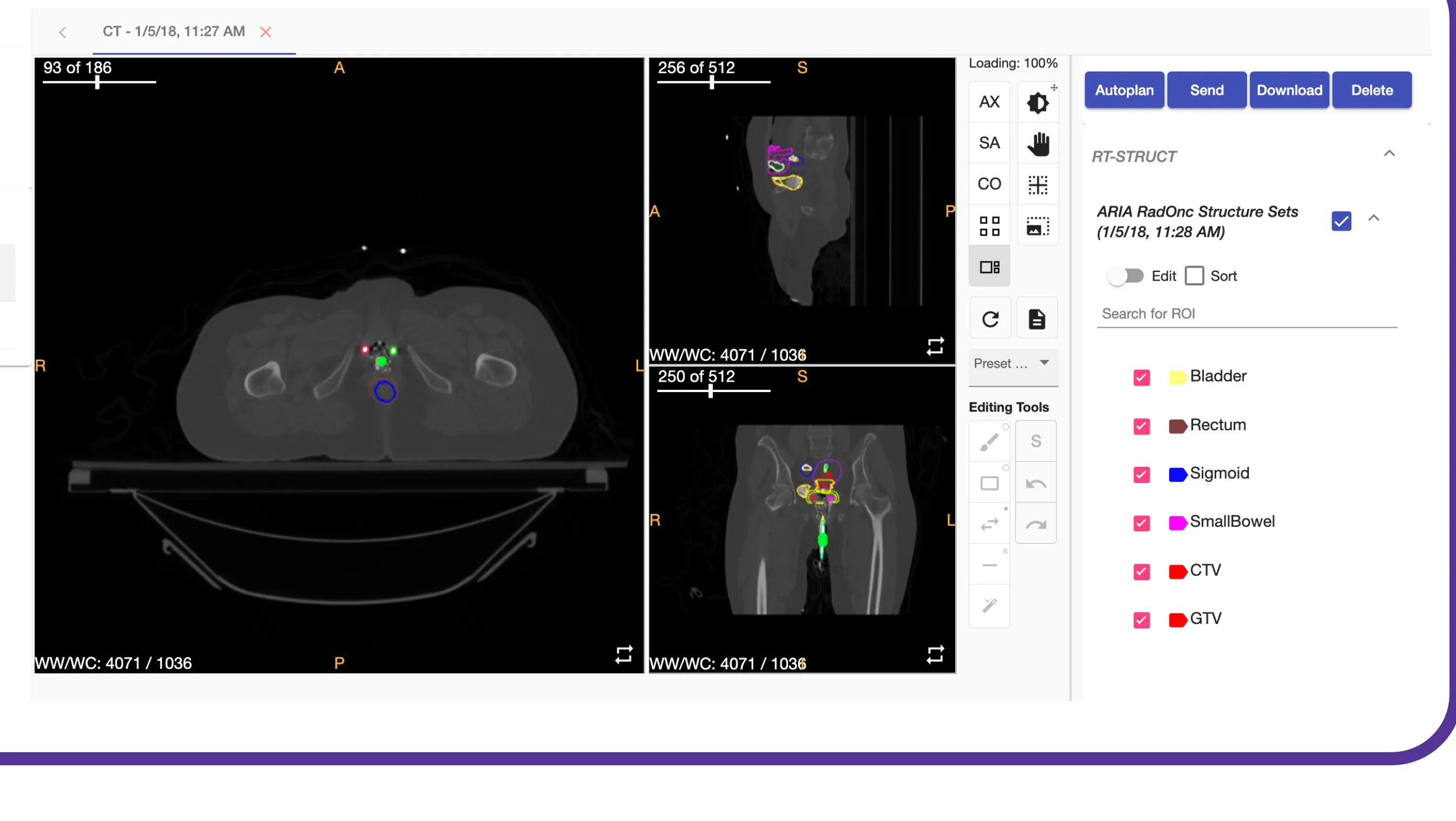
How it works
Four steps from CT to draft plan
Each step produces output you can inspect and edit — nothing is a black box between the planning CT and the plan.
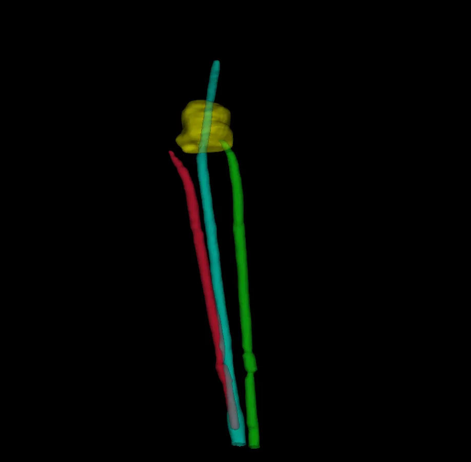
01 · Digitization — 3D geometry from the CT
- Applicator library. Pick cylinder, tandem-and-ring, tandem-and-ovoid, or interstitial; AutoBrachy digitizes the applicator in 3D from the planning CT.
- Needle identification. For interstitial cases, needles are identified and digitized alongside the applicator.
- Channel geometry for dose calculation. Digitized channels and dwell positions feed directly into optimization — no manual digitization pass.
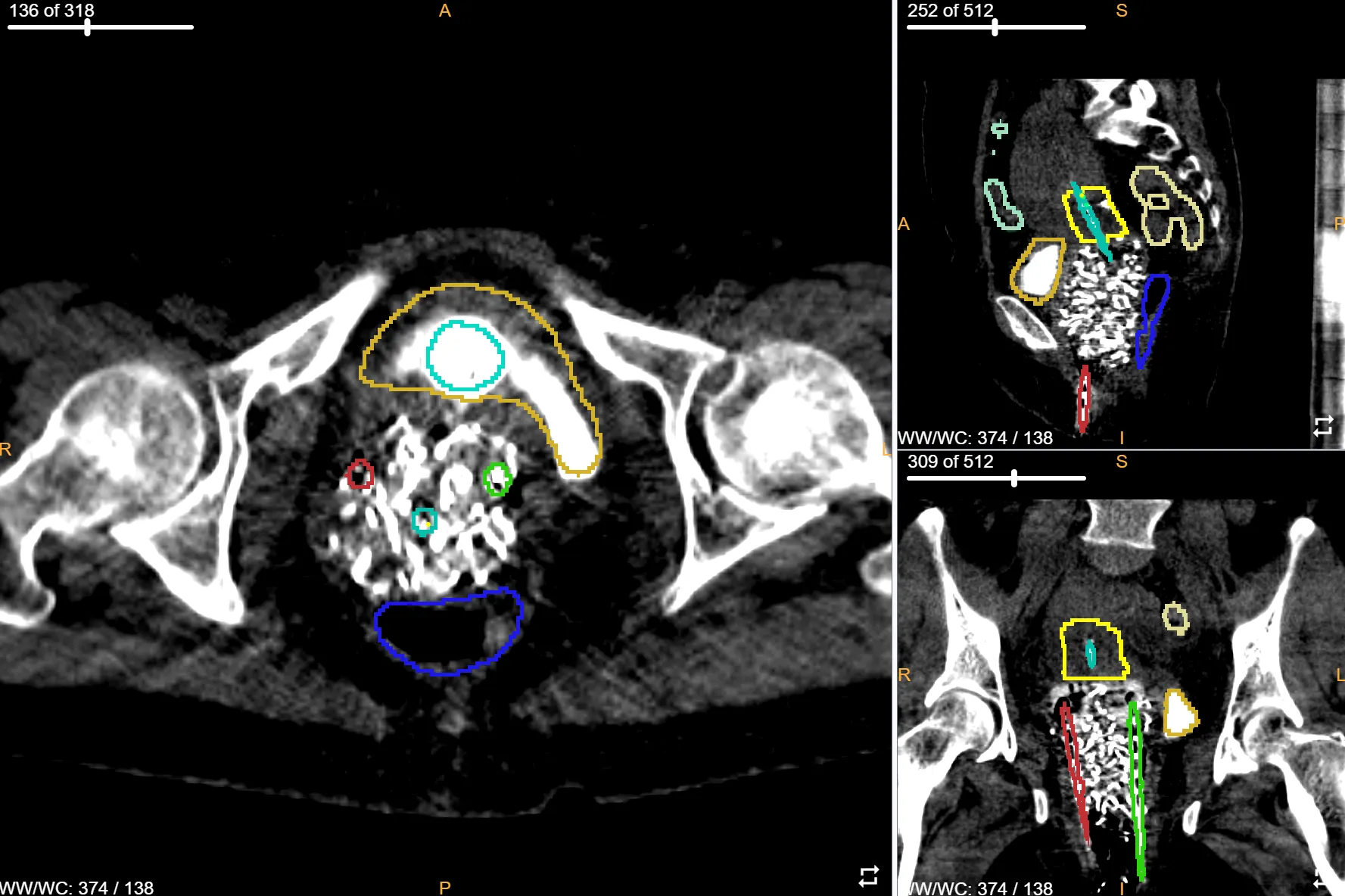
02 · Auto-contour — OARs + target
- Structures that matter for GYN HDR. Automatic contours for bladder, rectum, sigmoid, small bowel, and HR-CTV.
- Editable in place. Review and refine every contour in the same viewer before it feeds the plan — the physicist stays in control.
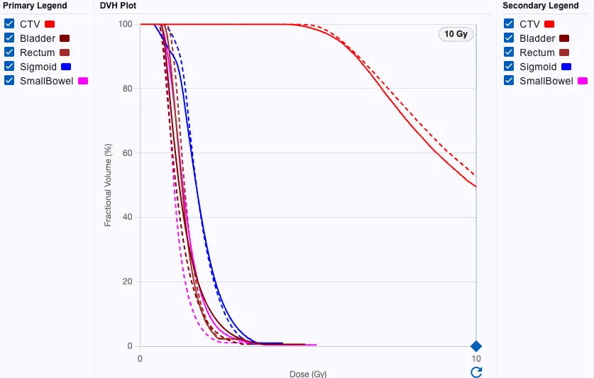
03 · Auto-plan — optimized dwell points + time
- Coverage and sparing. Protocol-driven optimization honors prescribed target coverage and OAR sparing for each structure.
- Interactive adjustment. Dose–volume histogram analysis and plan-adjustment tools for fine-tuning the draft before sign-off.
- Physicist-ready output. Isodose overlay, DVH, and dwell times — ready for review, edit, and clinician approval before treatment.
04 · Integrate — TPS round-trip via DICOM
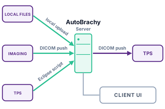
- Two ways to connect. Stand-alone web, or an Eclipse script that runs AutoBrachy without leaving the TPS.
- Standard DICOM round-trip. Approved plans return to your TPS as RTPLAN, RTSTRUCT, and RTDOSE — compatible with major TPS vendors.
Validation
Validated across institutions — and where the need is greatest
AutoBrachy's segmentation, applicator digitization, and dwell-time optimization have been evaluated on data the models never saw — at multiple institutions and in real low-resource clinics. Accepted for presentation at ABS and ASTRO 2026.
Multi-institutional validation
- Trained on one institution, tested on others. Models trained on 189 studies and externally validated on 202 more from an independent 54-patient cohort, spanning tandem-and-ovoid and tandem-and-ring cases the models never saw in training.
- Whole-pipeline evaluation. Automatic CTV and OAR segmentation (Dice and HD95), applicator digitization, and TG-43 dose calculation — assessed against expert contours and clinically delivered plans.
- Plan quality held or improved. CTV D90 matched the clinical plans, while OAR D2cc was comparable or significantly lower — better organ sparing — particularly for tandem-and-ovoid cases. Review remains recommended for sigmoid and small bowel.
Expanding access in low- and middle-income countries
- Real clinics, unseen anatomy. 14 tandem-and-ovoid cases from clinics in Uganda and Mongolia, planned by models trained only on U.S. expert data.
- About 95 minutes down to about 9. AutoBrachy produced complete 3D plans in roughly 9 minutes versus about 95 for manual planning — a ~90% reduction in end-to-end planning time.
- Better plans, not just faster. Improved HR-CTV D90 (8.35 vs 7.47 Gy) with lower OAR D2cc across bladder, rectum, sigmoid, and bowel — a path to leapfrog from 2D to 3D image-guided brachytherapy where expertise and time are scarce.
Presentations & manuscripts
Multi-Institutional Validation of an Automated Segmentation and Planning Reference System for Gynecologic HDR Brachytherapy — ABS 2026 · ASTRO 2026
AI-Based Brachytherapy Auto-Planning for Cervical Cancer in Low- and Middle-Income Countries — ASTRO 2026
Artificial Intelligence–Based Automated Brachytherapy Planning to Expand Access to 3D Image-Guided Treatment of Cervical Cancer in LMICs — manuscript in preparation
Technical specifications
What goes in, what comes out
Input
- DICOM CT images
- DICOM RT Structure Sets
- DICOM RT Plans
Output
- DICOM RT Structure Sets
- DICOM RT Plans
- DICOM RT Dose
- PDF plan report
Access
- Web-based interface — modern browser, no per-seat install
- Backend deploys locally or in the cloud
Deployment
Runs where your data lives
Deploy AutoBrachy locally inside your network or in the cloud. Patient data stays under your institution's control either way.
Scope & status
Where AutoBrachy is today
- Modality scope. GYN HDR brachytherapy — tandem-and-ring, tandem-and-ovoid, and interstitial applicators. Additional sites and LDR are on the roadmap.
- Regulatory status. AutoBrachy is an investigational device and is not FDA cleared. It is available for research use with our academic partners.
- Development partners & funding. Built and evaluated with medical physicists at UT Southwestern and UC San Diego, with SBIR funding from the National Cancer Institute.
See AutoBrachy on your own cases
AutoBrachy is investigational — not for clinical use.